New 3D Imaging Advancements for Heart Procedures
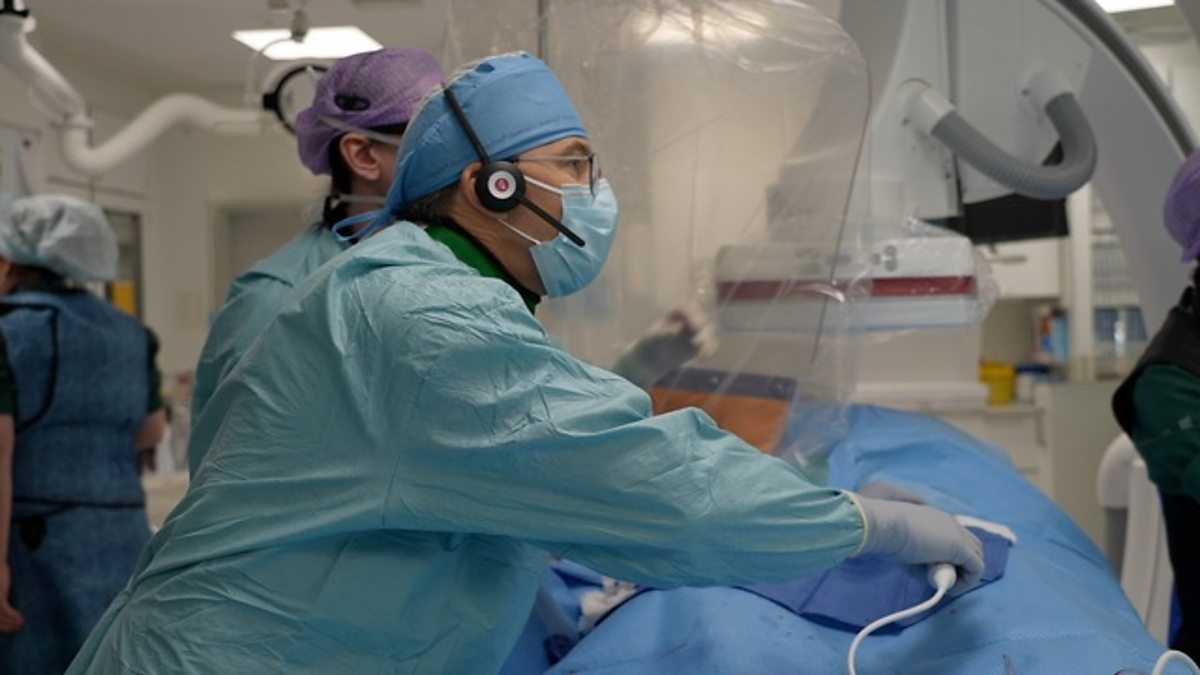
Learn how 3D spatial computing and AI are transforming high-stakes surgical precision in cardiology.
30-Second TL;DR
What Changed
Implementation of high-fidelity 3D imaging in cardiac surgery
Why It Matters
The adoption of AI-driven 3D reconstruction in medical imaging significantly reduces procedural risks. It sets a new standard for surgical planning and real-time guidance in cardiology.
What To Do Next
Explore medical imaging datasets on platforms like Kaggle or TCIA to understand how volumetric data is processed for AI-assisted diagnostics.
Key Points
- •Implementation of high-fidelity 3D imaging in cardiac surgery
- •Improved precision for surgeons during complex heart interventions
- •Integration of real-time visualization to enhance patient outcomes
Deep Insight
Background and context from public sources — not the original article. 31 sources cited.
Enhanced Key Takeaways
- •Augmented and Virtual Reality (AR/VR) technologies are increasingly utilized to provide immersive 3D environments for collaborative preoperative planning and real-time intraoperative overlays, enhancing surgeons' spatial understanding and precision beyond traditional 2D or static 3D displays.
- •Artificial intelligence (AI) is being integrated into 3D imaging workflows to automate the segmentation of CT and MRI images, enabling the rapid creation of patient-specific 3D heart models and improving the efficiency and accuracy of electroanatomic mapping systems for procedures like cardiac ablation.
- •Multimodality fusion imaging systems are emerging that combine real-time 2D fluoroscopy with pre-acquired 3D CT or MRI data, and integrate various imaging modalities such as ultrasound, CT, and angiography, to offer comprehensive and dynamic intraoperative guidance while potentially reducing radiation exposure for patients and staff.
- •Real-time 3D Transesophageal Echocardiography (3D TEE) has become a pivotal intraoperative tool for guiding a broad spectrum of percutaneous structural heart interventions, providing superior anatomical definition and immediate assessment of device positioning and function during procedures.
Competitor Analysis
- Key Features
- Integrated AI, real-time 2D/3D angiography, CT data integration, ultrasound monitoring
- Primary Application
- Coronary artery disease, heart valve interventions, stent implantation
- Unique Selling Proposition
- First-in-world clinical use of AI-integrated angiography system for precise treatment planning and reduced radiation.
- Key Features
- Holographic guidance, real-time 3D heart models, hand gesture manipulation, AR headset
- Primary Application
- Electrophysiology (EP) cardiac mapping, cardiac ablation
- Unique Selling Proposition
- Interactive 360-degree holographic visualization, allowing surgeons to manipulate images hands-free in a sterile field.
- Key Features
- Advanced electroanatomic mapping, high automation, hybrid impedance/magnetic field technology
- Primary Application
- Diagnosis and treatment of arrhythmias, cardiac ablation
- Unique Selling Proposition
- High precision and accuracy with automated point collection for rapid, high-density mapping.
- Key Features
- Electroanatomic mapping, magnetic tracking, 3D cardiac maps
- Primary Application
- Cardiac ablation, electrophysiology procedures
- Unique Selling Proposition
- Detailed 3D electroanatomic maps for visualizing and guiding catheter-based ablation.
- Key Features
- Non-invasive 3D cardiac maps, combines ECG signals with CT data, multi-chamber mapping
- Primary Application
- Characterizing abnormal heart rhythms, procedural planning outside EP lab
- Unique Selling Proposition
- Non-invasive mapping without catheterization, capturing rhythms across multiple chambers with a single beat.
- Key Features
- AI-driven automated segmentation of CT/MR images, digital twin of the heart
- Primary Application
- Image-guided ablations, pre-procedural planning
- Unique Selling Proposition
- Rapid creation of highly detailed, interactive patient-specific 3D heart models within hours using AI automation.
- Key Features
- Fuses 2D X-ray images with annotated pre-procedure 3D anatomical heart models (MRI/CT)
- Primary Application
- Cardiac biotherapeutic delivery, biopsy procedures, augmented fluoroscopy
- Unique Selling Proposition
- Enhances 2D fluoroscopy with precise 3D anatomical context for interventional cardiology.
| Company/System | Key Features | Primary Application | Unique Selling Proposition |
|---|---|---|---|
| Siemens Healthineers Artis Icono.vision | Integrated AI, real-time 2D/3D angiography, CT data integration, ultrasound monitoring | Coronary artery disease, heart valve interventions, stent implantation | First-in-world clinical use of AI-integrated angiography system for precise treatment planning and reduced radiation. |
| SentiAR CommandEP | Holographic guidance, real-time 3D heart models, hand gesture manipulation, AR headset | Electrophysiology (EP) cardiac mapping, cardiac ablation | Interactive 360-degree holographic visualization, allowing surgeons to manipulate images hands-free in a sterile field. |
| Abbott EnSite Precision Cardiac Mapping System | Advanced electroanatomic mapping, high automation, hybrid impedance/magnetic field technology | Diagnosis and treatment of arrhythmias, cardiac ablation | High precision and accuracy with automated point collection for rapid, high-density mapping. |
| Biosense Webster CARTO System | Electroanatomic mapping, magnetic tracking, 3D cardiac maps | Cardiac ablation, electrophysiology procedures | Detailed 3D electroanatomic maps for visualizing and guiding catheter-based ablation. |
| Medtronic CardioInsight Noninvasive 3D Mapping System | Non-invasive 3D cardiac maps, combines ECG signals with CT data, multi-chamber mapping | Characterizing abnormal heart rhythms, procedural planning outside EP lab | Non-invasive mapping without catheterization, capturing rhythms across multiple chambers with a single beat. |
| inHEART AI Software | AI-driven automated segmentation of CT/MR images, digital twin of the heart | Image-guided ablations, pre-procedural planning | Rapid creation of highly detailed, interactive patient-specific 3D heart models within hours using AI automation. |
| BioCardia/CART-Tech Heart3D Fusion Imaging | Fuses 2D X-ray images with annotated pre-procedure 3D anatomical heart models (MRI/CT) | Cardiac biotherapeutic delivery, biopsy procedures, augmented fluoroscopy | Enhances 2D fluoroscopy with precise 3D anatomical context for interventional cardiology. |
Technical Deep Dive
- 3D Transesophageal Echocardiography (3D TEE): Utilizes matrix array transducers for real-time 3D single-beat full volume acquisitions, often incorporating improved ultrasound quantification software. Features include multiplanar reformatting (MPR) for precise linear and annular measurements, and the ability to visualize structures from various 'surgical' en-face perspectives.
- Augmented Reality (AR) and Virtual Reality (VR) Systems: Involve head-mounted displays or spatial computing to project 3D virtual elements. These systems integrate imaging data from CT, MRI, 3D angiography, and 3D TEE, allowing for real-time overlay of virtual anatomical models onto the patient's body or immersive virtual environments for planning. Interaction often includes hand gestures to manipulate holographic models.
- Electroanatomic Mapping Systems (e.g., CARTO, EnSite): Employ either magnetic tracking (using sensors embedded in catheters and external magnetic field generators/location pads) or impedance-based electrofield mapping (using body-surface patch electrodes). Hybrid systems combine both. They construct real-time 3D cardiac maps by tracking catheter movement and electrical activity, with some advanced systems offering high-density mapping using multi-electrode catheters (e.g., 64-electrode mini-basket catheters).
- AI-Powered 3D Modeling Software (e.g., inHEART): Leverages artificial intelligence algorithms for fully automated segmentation of pre-procedural CT and/or MR images. This automation rapidly generates highly detailed, interactive 3D digital twins of the heart, optimizing pre-procedural planning for image-guided ablations.
- Fusion Imaging Systems: Integrate live 2D fluoroscopic images with pre-acquired 3D anatomical models (e.g., from CT or MRI). The 3D model is overlaid onto the real-time 2D image, providing enhanced anatomical context and guidance during interventional procedures.
- Robotic-Assisted Surgical Systems: Incorporate high-definition 3D imaging for visualization. Robotic instruments offer enhanced dexterity, allowing for rotations and curves not possible with human hands, facilitating minimally invasive procedures through small incisions.
Future ImplicationsAI analysis grounded in cited sources
Timeline
- 1990sInvention of 3D ultrasound prototype by Olaf von Ramm and Stephen Smith at Duke University.
- 2002Introduction of the first reasonably user-friendly real-time 3D echocardiography (RT3DE) for clinical use.
- 2011Development of a system for real-time 3D stereo visualization for cardiac ablation using 4D ultrasound and magnetic tracking.
- 2017Studies demonstrate improved surgical outcomes and reduced operating times using 3D-printed patient-specific heart models for preoperative planning.
- 2023-10SentiAR receives FDA 510(k) clearance for its CommandEP holographic guidance system for EP cardiac mapping.
- 2026-03Siemens Healthineers Artis Icono.vision system, an angiography system with integrated AI for real-time 2D/3D imaging, is introduced into clinical use.
Sources (31)
Factual claims are grounded in the sources below. Forward-looking analysis is AI-generated interpretation.
Weekly AI Recap
Read this week's curated digest of top AI events →
Related Updates


AI-curated news aggregator. All content rights belong to original publishers.
Original source: BBC Technology ↗
This is a summary, not the original. Read the source, or get the weekly briefing.
The weekly digest
One email a week. Unsubscribe anytime.