High schooler uses AI to screen autism via retina scans
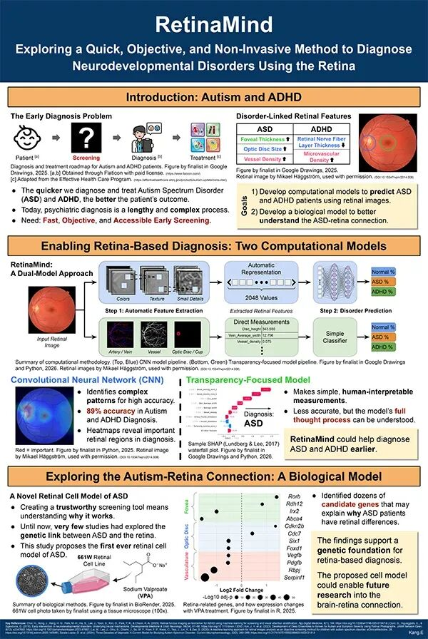
💡See how a high schooler used AI and CNNs to achieve 89% accuracy in screening for neurodevelopmental conditions.
⚡ 30-Second TL;DR
What Changed
RetinaMind uses convolutional neural networks to identify patterns in retinal images.
Why It Matters
This research bridges AI computation with biological science, offering a non-invasive, early-screening method for neurodevelopmental conditions. It emphasizes the importance of data diversity and clinical validation for AI-based medical tools.
What To Do Next
Explore the use of CNNs for medical imaging classification and investigate the integration of multi-modal data in diagnostic AI.
Key Points
- •RetinaMind uses convolutional neural networks to identify patterns in retinal images.
- •The tool achieved 89% accuracy in screening for autism and ADHD in test sets.
- •The project highlights the potential of using biological markers in the eye for neurological assessment.
🧠 Deep Insight
AI-generated analysis for this event — not the original article.
🔑 Enhanced Key Takeaways
- •The research leverages the 'retina-brain axis,' a biological concept suggesting that the retina acts as an extension of the central nervous system, allowing for non-invasive neurological observation.
- •The project was notably recognized at international science fairs, such as the Regeneron International Science and Engineering Fair (ISEF), which often serves as a launchpad for high-school-led medical AI innovations.
- •Unlike traditional diagnostic methods that rely on behavioral observation and lengthy questionnaires, this approach aims to reduce the average age of diagnosis, which currently remains high in many regions.
- •The study utilized publicly available retinal fundus image datasets, which were pre-processed to normalize lighting and contrast before being fed into the neural network.
- •Ethical considerations regarding the use of biometric retinal data for pediatric screening have been raised by medical experts, emphasizing the need for strict data privacy and informed consent protocols.
🛠️ Technical Deep Dive
- Architecture: Utilized a custom-trained Convolutional Neural Network (CNN) likely based on architectures such as ResNet or EfficientNet for feature extraction from fundus photography.
- Data Pre-processing: Images underwent CLAHE (Contrast Limited Adaptive Histogram Equalization) to enhance retinal vessel visibility and noise reduction.
- Input Data: High-resolution fundus images capturing the optic disc and macula regions.
- Training Methodology: Employed transfer learning techniques, fine-tuning pre-trained models on medical imaging datasets to overcome the limitation of a smaller sample size typical of student-led research.
🔮 Future ImplicationsAI analysis grounded in cited sources
⏳ Timeline
Weekly AI Recap
Read this week's curated digest of top AI events →
👉Related Updates



AI-curated news aggregator. All content rights belong to original publishers.
Original source: 虎嗅 ↗
This is a summary, not the original. Read the source, or get the weekly briefing.
The weekly digest
One email a week. Unsubscribe anytime.