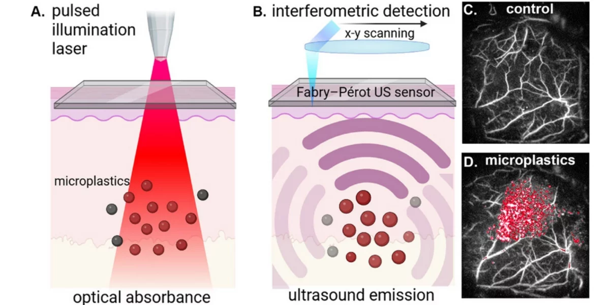
New Laser Imaging Tracks Microplastics in Living Tissue
💡A breakthrough in bio-imaging that opens new frontiers for AI-driven diagnostic and medical research applications.
⚡ 30-Second TL;DR
What Changed
Non-invasive laser imaging allows real-time tracking of microplastics in living organisms.
Why It Matters
This imaging advancement creates new datasets for computer vision and pattern recognition models in biological research.
What To Do Next
Explore computer vision libraries like OpenCV or PyTorch to process high-resolution biological imaging data for pattern detection.
🧠 Deep Insight
AI-generated analysis for this event.
🔑 Enhanced Key Takeaways
- •The imaging technique utilizes a method known as stimulated Raman scattering (SRS) microscopy, which detects the specific vibrational signatures of plastic polymers.
- •Researchers successfully tracked polystyrene microplastics as small as 200 nanometers, demonstrating high sensitivity to sub-micron particles.
- •The study revealed that microplastics can cross biological barriers, including the blood-brain barrier, which was previously difficult to visualize in vivo.
- •This imaging modality eliminates the need for fluorescent labeling, which often alters the physical properties or surface chemistry of the microplastics being studied.
- •The research team identified that particle size and surface charge significantly influence the rate and location of microplastic accumulation in organs like the liver and spleen.
🛠️ Technical Deep Dive
- Utilizes Stimulated Raman Scattering (SRS) microscopy to achieve label-free chemical imaging.
- Employs dual-beam laser excitation to target the characteristic C-H stretching vibrations of polymer chains.
- Capable of deep-tissue penetration by utilizing near-infrared (NIR) laser wavelengths to minimize scattering and phototoxicity.
- Integrates high-speed scanning galvo-mirrors to enable real-time, video-rate acquisition of microplastic movement.
- Employs computational spectral unmixing algorithms to differentiate microplastic signals from endogenous biological molecules like lipids and proteins.
🔮 Future ImplicationsAI analysis grounded in cited sources
⏳ Timeline
Weekly AI Recap
Read this week's curated digest of top AI events →
👉Related Updates
Same topic
Explore #bio-imaging
Same product
More on laser-imaging-technology
Same source
Latest from cnBeta (Full RSS)
Building a Flow Matching Image Generator from Scratch
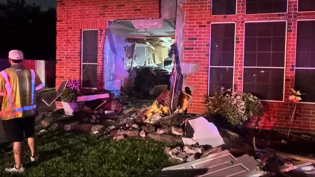
Tesla FSD Under Scrutiny After Fatal Texas Crash

Physical Media Declining as Digital Codes Become Industry Norm
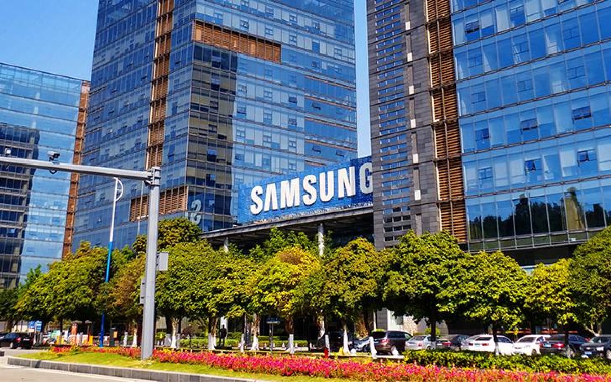
Anthropic in talks with Samsung for custom AI chips
AI-curated news aggregator. All content rights belong to original publishers.
Original source: cnBeta (Full RSS) ↗