AI Helps Surgeons Remove Brain Tumour Safely
💡See how real-time surgical vision AI guided a landmark brain tumour operation.
⚡ 30-Second TL;DR
What Changed
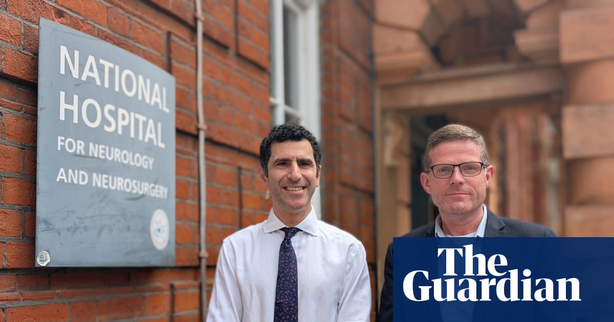
The operation took place in May at the National Hospital for Neurology and Neurosurgery in London.
Why It Matters
Real-time surgical AI could reduce the risk of damaging vital structures and provide clinicians with an additional layer of visual guidance. However, broader adoption will require extensive clinical validation, regulatory approval, explainability, and robust safeguards against missed or incorrect anatomy detection.
What To Do Next
Prototype a computer-vision pipeline for surgical video segmentation, then benchmark anatomy-detection accuracy and latency on de-identified clinical footage before considering clinical use.
Key Points
- •The operation took place in May at the National Hospital for Neurology and Neurosurgery in London.
- •AI analyzed live camera footage during surgery to identify critical anatomy that surgeons needed to avoid.
- •The procedure reportedly saved the sight of 48-year-old patient Rhys Hibbert.
- •The milestone highlights a potential role for computer vision in high-risk, precision surgery.
🧠 Deep Insight
Background and context from public sources — not the original article. 8 sources cited.
🔑 Enhanced Key Takeaways
- •The AI system was trained on a dataset comprising hundreds of historical surgical videos to achieve the pattern recognition necessary for identifying complex intracranial structures.
- •The specific tumor targeted in the procedure was an 11mm growth located on the patient's pituitary gland, which posed a direct threat to the optic chiasm.
- •Post-operative recovery was rapid, with the patient achieving independent mobility within seven days and reporting a restored 360-degree panoramic field of vision.
- •The technology utilizes real-time segmentation to overlay color-coded visual cues onto the surgeon's field of view, distinguishing between healthy tissue, blood vessels, and nerves.
- •The broader field of AI-assisted neuro-oncology is seeing increased capital inflow, evidenced by a £100,000 investment in August 2026 into Neurolase, a firm specializing in tissue-differentiation software.
📊 Competitor Analysis▸ Show
| Feature | SurgAR | Neurolase | NHNN AI System |
|---|---|---|---|
| Primary Focus | Augmented Reality/Digital Twins | Tissue differentiation | Real-time surgical navigation |
| Development Stage | Clinical/Research | Early-stage investment | First-in-human successful use |
| Key Capability | Hidden tumor visualization | Cancerous vs. healthy tissue mapping | Critical anatomy segmentation |
🛠️ Technical Deep Dive
- The system employs computer vision algorithms to perform real-time segmentation of surgical video feeds.
- The architecture relies on deep learning models trained on large-scale historical surgical video archives to identify anatomical landmarks.
- The implementation involves overlaying visual markers on the surgical display to provide color-coded guidance for blood vessels and nerves.
- The platform integrates with existing surgical camera hardware to provide low-latency feedback during the procedure.
🔮 Future ImplicationsAI analysis grounded in cited sources
⏳ Timeline
📎 Sources (8)
Factual claims are grounded in the sources below. Forward-looking analysis is AI-generated interpretation.
Weekly AI Recap
Read this week's curated digest of top AI events →
👉Related Updates
AI-curated news aggregator. All content rights belong to original publishers.
Original source: The Guardian Technology ↗
This is a summary, not the original. Read the source, or get the weekly briefing.
Weekly AI briefing
One email a week. Unsubscribe anytime.